
 "Hidden universe captured in astonishing detail by winners of Nikon micro-photography contest")
As fascinating as the world looks through human eyes, there’s an entire world invisible to our naked eyes. The microscope lends us access to see, even interact with the wonder, beauty and breathtaking diversity in the microscopic universe. Each year, the Nikon Small World contest chooses a series of photographs taken through the year of the microscopic universe. In the 45th year of the ‘Small World’ competition, a panel of microscopy experts and science journalists judged and reviewed over 2,000 images from individuals and institutions across a hundred countries to gather the finest of microphotography in 2019. The images submitted range across the sciences — from tiny lumps of crystallised copper oxide with its harsh facades and jutting angles to the intricate nerves and skeletal system of an alligator embryo. It was the blown-up biology that won the day, with stunning images of embryos, cells, plants, organs and animals. Here are the 20 winning images from this year’s Nikon Small World photography contest.
1st place winner: Flourescent turtle embryo
[caption id=“attachment_7534611” align=“alignnone” width=“1280”] 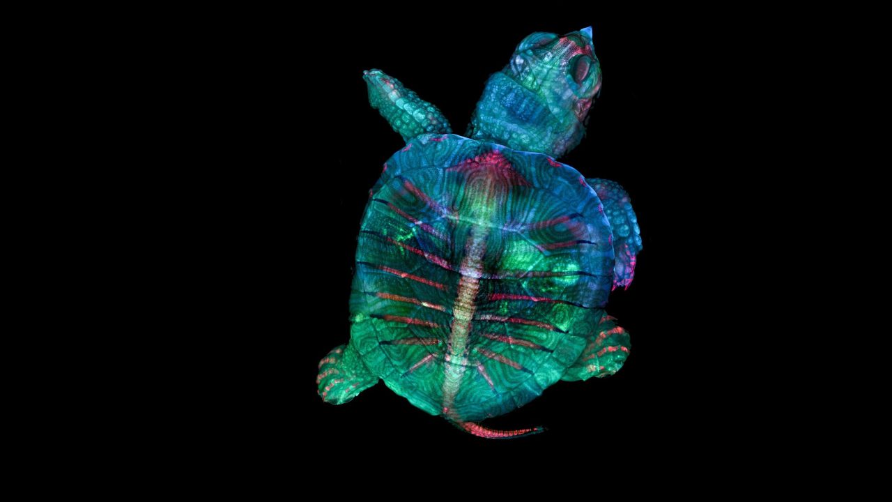 1st place: Fluorescent turtle embryo. Image credit: Teresa Zgoda and Teresa Kugler/Nikon Small World[/caption]
2nd place winner: Single-cell, freshwater protozoans
[caption id=“attachment_7534261” align=“alignnone” width=“1484”] 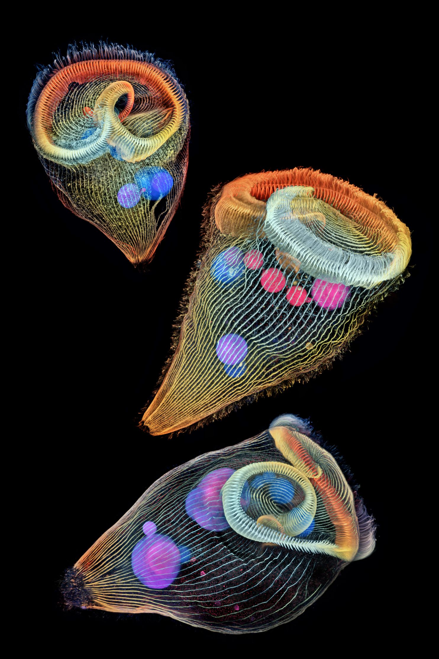 Depth-colour-coded projection of three stentors (single-cell freshwater protozoans). Image credit: Dr Igor Siwanowicz/Nikon Small World[/caption]
3rd place winner**: Alligator embryo developing nerves and skeleton**
[caption id=“attachment_7534751” align=“alignnone” width=“1280”]  In third place, this photo of an alligator embryo developing nerves and skeleton. Image credit: Daniel Smith Paredes & Dr Bhart Anjan S Bhullar/Nikon Small World[/caption]
4th place winner**:** A male mosquito
[caption id=“attachment_7534721” align=“alignnone” width=“1280”] A male mosquito. Image credit: Jan Rosenboom/Nikon Small World[/caption]
5th place winner: The perfect snowflake
[caption id=“attachment_7534841” align=“alignnone” width=“1280”]  All snowflakes have six sides, like this one picture in the 5th-place-winning photo at four times magnification. Image credit: Caleb Foster/Nikon Small World[/caption]
6th place winner:
[caption id=“attachment_7534851” align=“alignnone” width=“1280”] At 6th place is this image of a small white-hair spider. Image credit: Javier Ruperez/Nikon Small World[/caption]
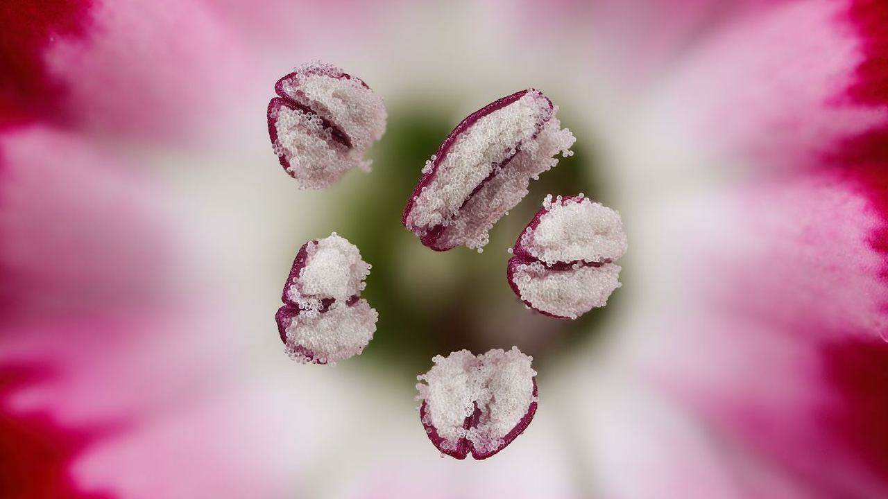
7th place winner: Chinese red carnation stamen
[caption id=“attachment_7534921” align=“alignnone” width=“1280”] The photograph shows Chinese red carnation stamen in 3X. Image credit: Dr Guillermo Lopez Lopez/Nikon Small World[/caption]
8th place winner: A frozen water droplet
[caption id=“attachment_7534971” align=“alignnone” width=“1280”] A frozen water droplet under 8X magnification won 8th place. Image credit: Garzon Christian/Nikon Small World[/caption]
9th place winner: Tulip bud cross-section
[caption id=“attachment_7535131” align=“alignnone” width=“1280”] This photograph of a tulip bud cross-section stood 9th place this year. Image credit: Andrei Savitsky and Cherkassy/Nikon Small World[/caption]
10th place winner: A cell mid-division
[caption id=“attachment_7535201” align=“alignnone” width=“1280”] BPAE (Bovine Pulmonary Artery Endothelial) cells in the telophase stage of mitosis (cell division). Image credit: Jason M Kirk/Nikon Small World[/caption]
11th place winner: Fruit fly ovaries
[caption id=“attachment_7535261” align=“alignnone” width=“1280”] 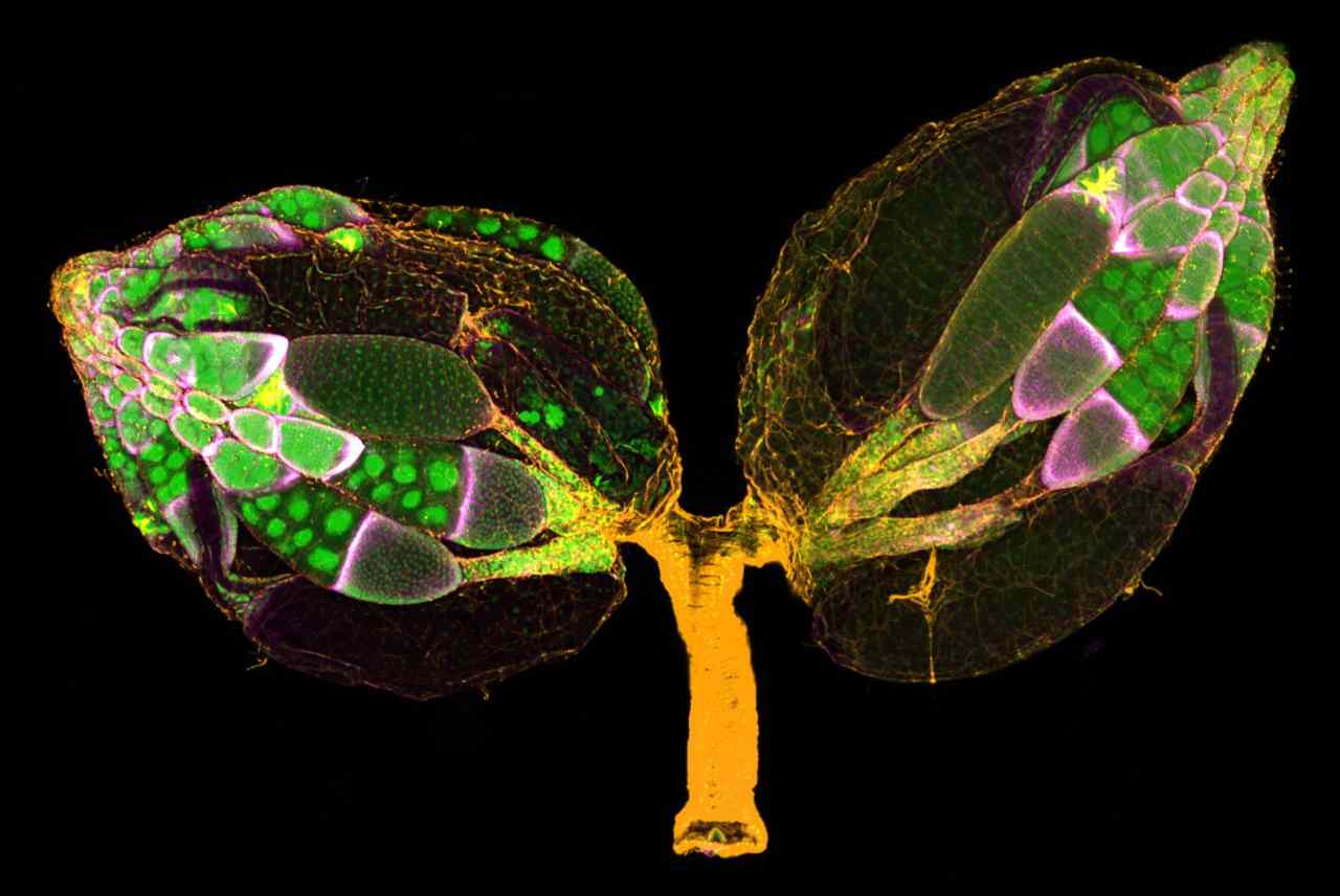 At 11th place is this vivid snap of a pair of ovaries from an adult Drosophila (fruit fly). Image credit: Dr Yujun Chen & Dr Jocelyn McDonald/Nikon Small World[/caption]
12th place winner: Mosquito larva
[caption id=“attachment_7535281” align=“alignnone” width=“1280”] 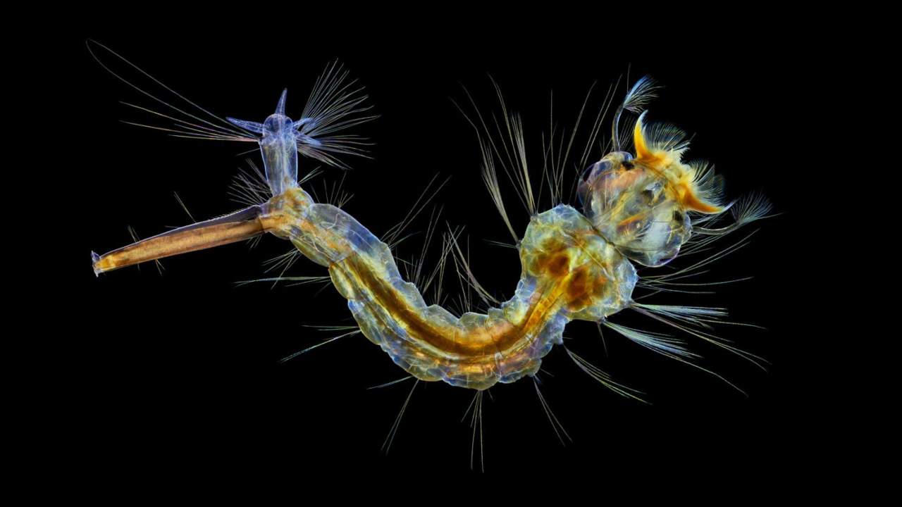 This image of a mosquito larva stood 12th overall. Image credit: Anne Algar/Nikon Small World[/caption]
13th place winner: A microscopic cuprite castle
[caption id=“attachment_7535331” align=“alignnone” width=“1280”] 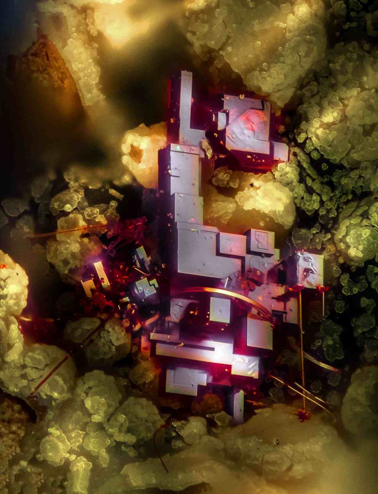 Cuprite, a mineral composed of copper oxide under 20X magnification. Image credit: Dr Emilio Carabajal Márquez/Nikon Small World[/caption]
14th place winner: The female lynx spider
[caption id=“attachment_7535391” align=“alignnone” width=“1280”] 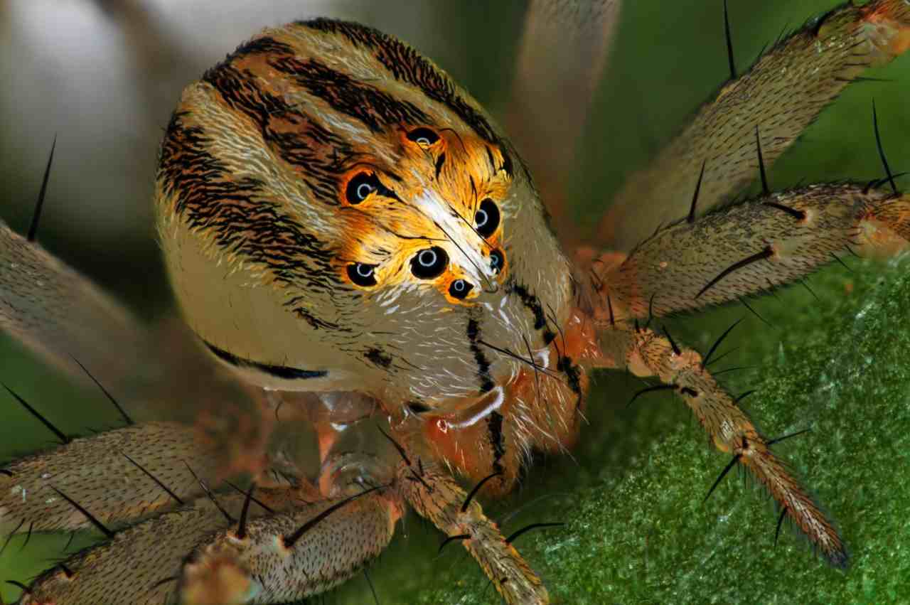 This portrait of the Oxyopes dumonti (lynx) spider female clinched the 14th place. Image credit: Antoine Franck-Cirad/Nikon Small World[/caption]
15th place winner: A pregnant Daphnia magna
[caption id=“attachment_7535481” align=“alignnone” width=“1280”] 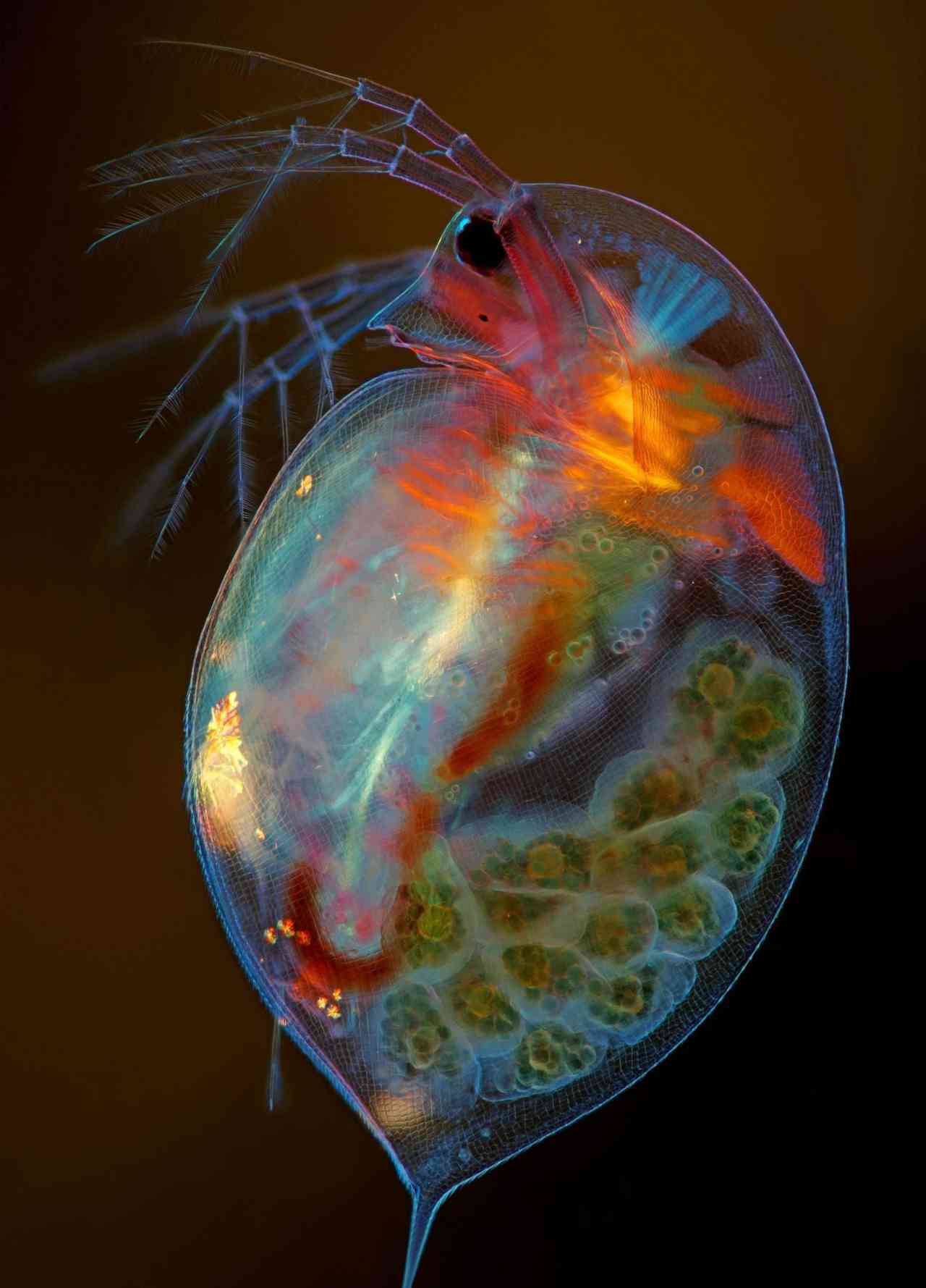 At 15th place, a pregnant, small plankton (crustacean) from the Daphnia magna species. Image credit: Marek Mis/Nikon Small World[/caption]
16th place winner: Compound eye of a housefly
[caption id=“attachment_7535541” align=“alignnone” width=“1280”] 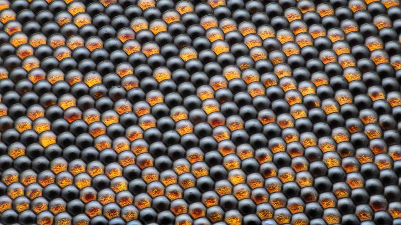 This 50X image of a housefly’s compound eye pattern won 16th place in the contest overall. Image credit: Dr Razvan Cornel Constantin/Nikon Small World[/caption]
17th place winner: Vitamin C
[caption id=“attachment_7535581” align=“alignnone” width=“1280”] 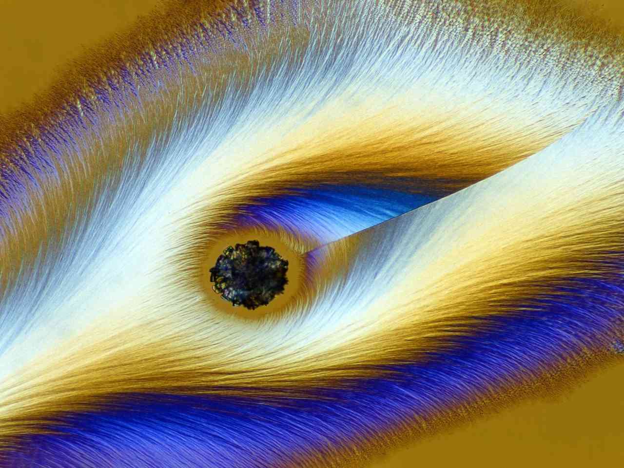 This image at 4X magnification of Vitamin C under the microscope won 17th position in the contest. Image credit: Karl Deckart/Nikon Small World[/caption]
18th place winner: a Cristobalite crystal in quartz
[caption id=“attachment_7535601” align=“alignnone” width=“1280”] 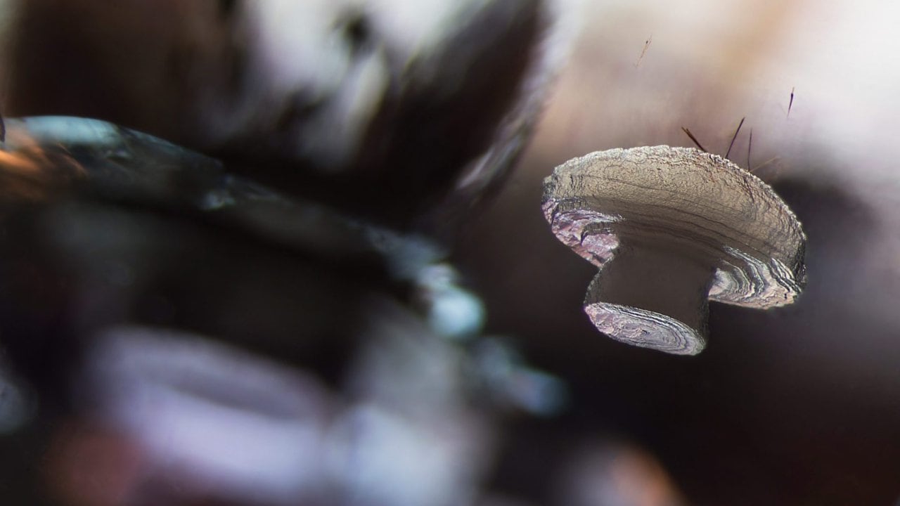 A crystal of Cristobalite suspended in a quartz mineral host clinched the 18th position. Image credit: E Billie Hughes/Nikon Small World[/caption]
19th place winner: Octopus embryos
[caption id=“attachment_7535621” align=“alignnone” width=“1280”]  A fluorescent octopus bimaculoides embryo, as seen under 5X magnification won 19th place. Image credit: Martyna Lukoseviciute, Dr Carrie Albertin/Nikon Small World[/caption]
20th place winner: Mouse heart and blood vessels
[caption id=“attachment_7535651” align=“alignnone” width=“1280”] In 20th place, blood vessels of a mouse heart following a heart attack. Image credit: Simon Merz Lea Bornemann, Sebastian Korste/Nikon Small World[/caption]
 "Israel targets top Hamas leaders in Doha; Qatar, Iran condemn strike as violation of sovereignty")
 "Nepal: Oli to continue until new PM is sworn in, nation on edge as all branches of govt torched")
 "Who is CP Radhakrishnan, India's next vice-president?")
 "Israel informed US ahead of strikes on Hamas leaders in Doha, says White House")


](https://images.firstpost.com/wp-content/uploads/large_file_plugin/2019/10/1571721898_1stplace_Fluorescentturtleembryo_TeresaZgodaandTeresaKugler_NikonSmallWorld.jpg)
](https://images.firstpost.com/wp-content/uploads/large_file_plugin/2019/10/1571721916_2ndplace_Depth-colorcodedprojectionsofthreestentorssingle-cellfreshwaterprotozoans_DrIgorSiwanowicz_NikonSmallWorld.jpg)
](https://images.firstpost.com/wp-content/uploads/large_file_plugin/2019/10/1571721962_3rdplace_Alligatorembryodevelopingnervesandskeleton_DanielSmithParedes&DrBhartAnjanSBhullar_NikonSmallWorld.jpg)
](https://images.firstpost.com/wp-content/uploads/large_file_plugin/2019/10/1571722000_5thplace_Allsnowflakeshavesixsides,likethisonepicturedhereatfourtimesmagnification_CalebFoster_NikonSmallWorld.jpg)
](https://images.firstpost.com/wp-content/uploads/large_file_plugin/2019/10/1571722161_11thplace_OvariesfromanadultDrosophilaorfruitfly_DrYujunChen&DrJocelynMcDonald_NikonSmallWorld.jpg)
](https://images.firstpost.com/wp-content/uploads/large_file_plugin/2019/10/1571722182_12thplace_Mosquitolarva_AnneAlgar_NikonSmallWorld.jpg)
](https://images.firstpost.com/wp-content/uploads/large_file_plugin/2019/10/1571722213_13thplace_CupritemineralcomposedofcopperoxideDrEmilioCarabajalM%c3%a1rquez_NikonSmallWorld.jpg)
](https://images.firstpost.com/wp-content/uploads/large_file_plugin/2019/10/1571722823_14thplace_OxyopesdumontifemelleAntoineFranck-Cirad_NikonSmallWorld.jpg)
](https://images.firstpost.com/wp-content/uploads/large_file_plugin/2019/10/1571722848_15thplace_PregnantDaphniamagnasmallplanktoniccrustacean_MarekMis_NikonSmallWorld.jpg)
](https://images.firstpost.com/wp-content/uploads/large_file_plugin/2019/10/1571722887_16thplace_Houseflycompoundeyepattern_DrRazvanCornelConstantin_NikonSmallWorld.jpg)
](https://images.firstpost.com/wp-content/uploads/large_file_plugin/2019/10/1571722913_17thplace_VitaminC_KarlDeckart_NikonSmallWorld.jpg)
](https://images.firstpost.com/wp-content/uploads/large_file_plugin/2019/10/1571722934_18thPlace_Cristobalitecrystalsuspendedinitsquartzmineralhost_EBillieHughes_NikonSmallWorld.jpg)
](https://images.firstpost.com/wp-content/uploads/large_file_plugin/2019/10/1571722947_19thplace_Aflourescentoctopusbimaculoidesembryo_MartynaLukoseviciuteDrCarrieAlbertin_NikonSmallWorld.jpg)