
The miniature marvels of the world unlocked!
Few would be able to guess what these images are, but that’s the beauty of microscopic photography. From slime mold to milk-producing cells, the winners of this year’s Nikon Small World photo contest reveal the stunning sights of our universe — under a microscope!
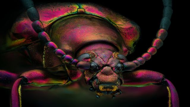)
Image of distinction: A red speckled jewel beetle (Chrysochroa buqueti rugicollis) magnified three times. Image Courtesy: Yousef Al Habshi
)
First place winner: An embryonic hand of a Madagascar giant day gecko. Image Courtesy: Grigorii Timin, Dr Michel Milinkovitch
)
Second place winner: Breast tissue showing contractile myoepithelial cells wrapped around milk-producing alveoli. Image Courtesy: Dr Caleb Dawson
)
Image of distinction: A dental drill bit studded with diamond chips. Image Courtesy: Karl E Deckart
)
Image of distinction: What butterfly scales look like under the magnifying glass. Image Courtesy: Yuan Ji
)
Image of distinction: A winged ant encased in approximately 20 million-year-old Dominican amber. Image Courtesy: Enrico Bonino
)
Tenth place: A magnified view of a fly under the chin of a tiger beetle. Image Courtesy: Murat Öztürk
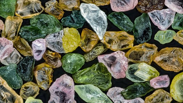)
Image of distinction: Grains of sand from Alaska. Image Courtesy: Xinpei Zhang
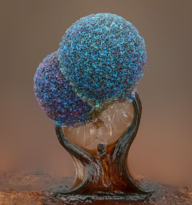)
Fifth place: This closeup view of slime mold on a leaf is magnified 10 times normal size. Most slime molds have smooth heads but this pair features what look like wrinkled pompoms. Image Courtesy: Alison Pollack
)
Sixth place: Unburned pieces of carbon drift away from an extinguished candle in this photograph, magnified 2.5 times normal size. Image Courtesy: Ole Bielfeldt
)
Image of distinction: A moss spore capsule. Image Courtesy: Michael Landgrebe
)
Seventeenth place: A tail fin of a zebrafish larva with peripheral nerves (green) and extracellular matrix (violet). Image Courtesy: Dr Daniel Wehner & Julia Kolb
 "‘I’d love to do it’: Trump refuses to rule out third term — despite constitutional limit")
 "What is the ‘Golden Fleet’ of navy ships that Trump wants to counter China with?")
 "‘Ramayan country’: Why Trinidad and Tobago wants to build a large Ram temple")
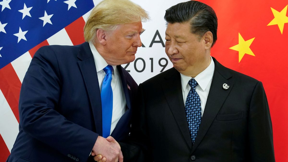 "How China's rare earths choke changed America's trade deals")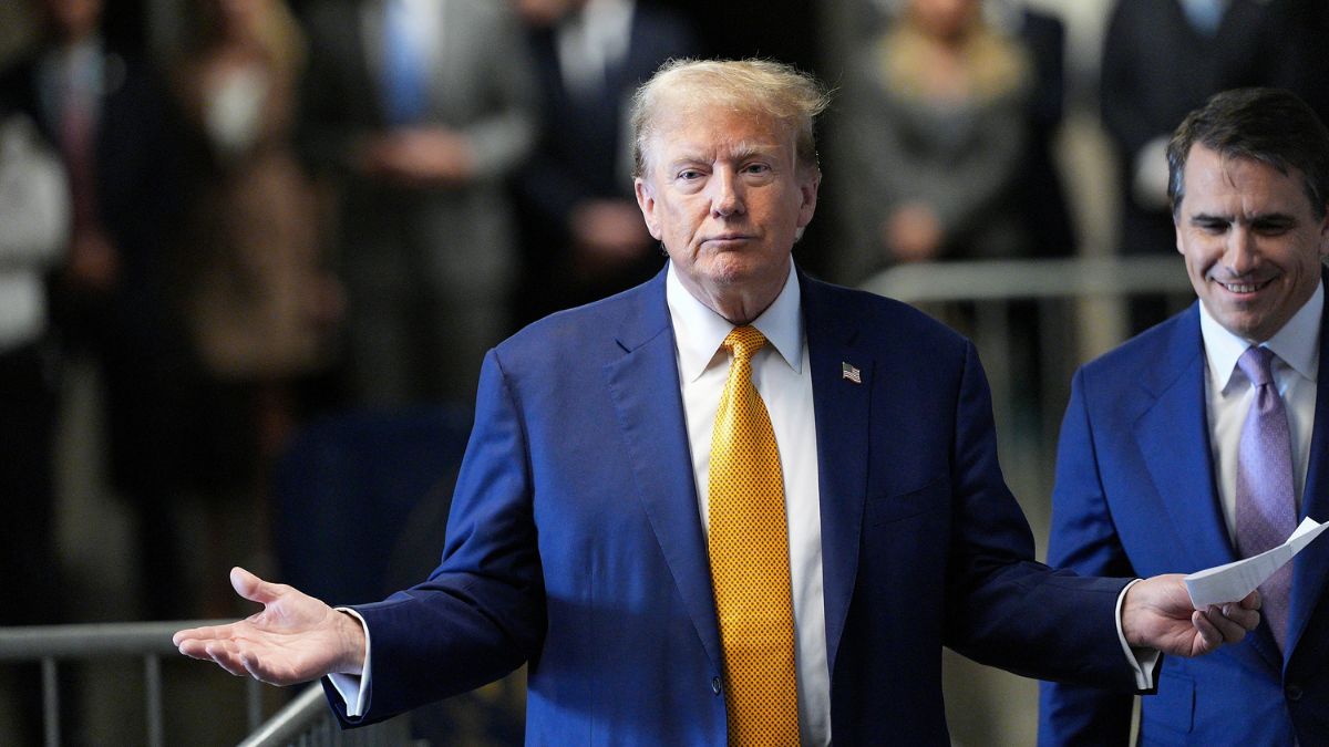
Top Shows


