
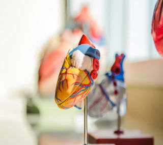
 "Scientists have managed to take a 3D bioprint of a human heart, a significant step with the potential to save many lives")
3D bioprinting may be a $4.1 billion market by 2026, according to US-based market research company Grand View Research, Inc. In mid-April, Israeli scientists bioprinted a tiny human heart complete with cells, blood vessels, ventricles and chambers. Strides like these could make organ waitlists a thing of the past. So, what is 3D bioprinting? And if it continues to grow at this clip, what are its implications for human health and heart disease in India? [caption id=“attachment_7276931” align=“alignleft” width=“320”]  Representative image. Image by StockSnap from Pixabay[/caption] How did we get here? In 1983, Charles Hull 3D-printed a small, black eye-wash cup: arguably the first 3D printed object in the world. In 1986, he patented his technology stereolithography and founded 3D Systems in South Carolina, US. Just 13 years later, in 1996, surgeons at the Texas, US-based Wilford Hall medical centre, used technology made by Hull’s company to create a model of conjoined twins. The purpose: to come up with strategies to separate them. In the end, the surgeons were able to do this so successfully that each twin was able to walk after the procedure. Scientists have since used 3D bioprinting to make stents and splints that are now commercially viable. In 2002, scientists were able to bioprint a miniature kidney. Now, just 17 years later, scientists have managed to print a human heart. Albeit the size of a bunny’s heart, it is a big step with big potential and a big thumbs up for human ingenuity. How does it work? Freeform Reversible Embedding of Suspended Hydrogels or FRESH as the most popular technique for bioprinting is called, basically works off 3D imaging: MRIs or CT scans produce 3D images of the organ to be printed. Next, scientists use biological materials like alginate, collagen, gelatin, hyaluronic acid and decellularized extracellular matrix scaffolds to ‘print’ the organ. The bioprinted heart is evidence of the level of complexity that scientists are able to achieve with this process. Sean V. Murphy and Anthony Atala explained this beautifully in ‘3D bioprinting of tissues and organs’, an article published in the peer-reviewed journal Nature Biotechnology, in August 2014. “In 3D bioprinting, layer-by-layer precise positioning of biological materials, biochemicals and living cells, with spatial control of the placement of functional components, is used to fabricate 3D structures. There are several approaches to 3D bioprinting, including biomimicry, autonomous self-assembly and mini-tissue building blocks,” they wrote. Biomimicry uses a minute understanding of the micro-environment of an organ to copy or recreate the exact cellular and extracellular components of the organ. Autonomous self-assembly does not use a scaffold. Instead, it learns from embryonic organ development, to make cellular spheroids (the biological material here) that assemble and fuse to form the desired organ. Finally, the mini-tissue approach breaks each organ down to minuscule sub-parts and prints these out. These ‘building blocks’ are then brought together through self-assembly or by implementing a ‘rational design’. In each of these methods, scientists use a variety of biological materials. 3D bioprinting uses bioink to ‘print out’ new tissue. This bioink is made up of living cells and biopolymer gels which act as molecular scaffolds. The biopolymer gels rapidly diffuse into the solution and form networks through crosslinking - these networks eventually form the core structure of the organ. To make a living organ, it is important to infuse it with the inherent cellular environment. For this, scientists take hematopoietic stem cells (stem cells that make blood cells) from the patient’s blood and allow them to form the different lineages of blood cells. By depositing this bioink layer-by-layer, scientists can print out the whole organ. What does the future hold? 3D bioprinting has tremendous potential for regenerative medicine. We are already able to successfully culture stem cells and use them to engineer human tissue. We can now repair certain types of nerve damage with the help of this technology. In January 2018, 3D bioprinting methods came to a 22-year-old woman’s rescue in an unusual way. When her doctors in Dubai screened her father — who was to donate the kidney for her transplant — they found a cancerous cyst on his kidney. Naturally, they didn’t do the transplant. In future, ‘printing out’ entire organs, complete with complex vascular systems will make replacement organs available to millions of patients on waitlists for kidney transplant, heart transplant, liver transplant and lung transplant. Not only this, specially printed replacement organs will be a perfect match for the organ receiver. This means transplant patients will now be free from some of the complications associated with a donor organ. Rejection rate will come down. Advanced 3D bioprinting will also enable the regeneration of new body parts and model organs. In April 2019, scientists N. Sigaux, L. Pourchet, P. Bretona and S. Brosset et al., wrote in the Journal of Stomatology, Oral and Maxillofacial Surgery: “Once the barrier of vascularization is overcome, printing organs and composite tissues of any size could be possible, opening the doors for personalized medicine based on medical imaging. Printing custom-made autologous grafts or flaps could minimize donor site morbidity and maximize the morphological results.” What are the challenges? Maintaining the shape of the organ while also maintaining the surrounding environment to allow the organ to grow and mature is a particularly big hurdle. The solution — to build an appropriate bioink — is easier said than done. 3D bioprinting has tremendous potential for organ transplants - on paper. In reality, scientists are yet to test its viability inside the human body. The scientists at Tel Aviv University overcame some of these challenges to bioprint a complete human heart. And that’s why, in mid-April, they caught our attention in India and across the world. Once scientists are able to print life-size organs, it could transform medicine as we know it. Health articles in Firstpost are written by myUpchar.com, India’s first and biggest resource for verified medical information. At myUpchar, researchers and journalists work with doctors to bring you information on all things health. To know more on this topic, please visit https://www.myupchar.com/en/disease/heart-transplant
3D bioprinting has huge potential for regenerative medicine. We are already able to successfully culture stem cells and use them to engineer human tissue.
Advertisement
End of Article
 "Israel targets top Hamas leaders in Doha; Qatar, Iran condemn strike as violation of sovereignty")
 "Nepal: Oli to continue until new PM is sworn in, nation on edge as all branches of govt torched")
 "Who is CP Radhakrishnan, India's next vice-president?")
 "Israel informed US ahead of strikes on Hamas leaders in Doha, says White House")


](https://images.firstpost.com/wp-content/uploads/2019/09/heart-2607178_1280_.jpg)